Are you a dentist struggling to consistently obtain high-quality dental x-ray images? Poor image quality can significantly hinder accurate diagnosis, leading to delayed treatment and potentially compromised patient outcomes. The ability to interpret radiographic images accurately depends heavily on their clarity and detail. This comprehensive guide delves into the critical factors influencing image quality in dental radiography and provides practical strategies for achieving superior results – ultimately empowering you to make more confident diagnoses and deliver exceptional care.
Introduction: The Foundation of Accurate Diagnosis
Dental x-rays are an indispensable tool in modern dentistry, providing a window into the hidden structures within the mouth. However, simply taking an image isn’t enough; the quality of that image directly impacts the ability to detect subtle abnormalities like caries (tooth decay), periodontal disease (gum disease), and bone loss. A blurry or obscured x-ray can lead to missed diagnoses, requiring further investigation and potentially delaying effective treatment. Early detection is key when it comes to oral health, and high-quality images are the cornerstone of this process.
The pursuit of optimal image quality in dental radiography isn’t merely about technical perfection; it’s about patient care. Clearer images translate directly into more confident diagnoses and better treatment plans. This article will explore various factors contributing to image quality, from equipment selection and technique execution to post-processing strategies. We’ll examine how advancements in technology like cone beam computed tomography (CBCT) are revolutionizing dental imaging and discuss best practices for maximizing the diagnostic value of every x-ray.
Factors Affecting Image Quality in Dental X-Rays
1. Equipment – The Starting Point
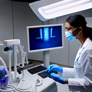
The equipment used to generate dental x-rays plays a fundamental role in image quality. Different types of imaging systems offer varying levels of resolution and contrast. Digital radiography (DR), which uses sensors to capture images electronically, is now the dominant technology due to its speed, reduced radiation exposure, and superior image characteristics compared to traditional film radiography.
- Film Radiography: While largely replaced, film systems still require precise technique and careful processing. Film quality significantly impacts the final image.
- Digital Radiography (DR): Offers immediate images, eliminating the need for chemical development. DR sensors are sensitive to X-ray energy, resulting in sharper images with better contrast. The type of sensor (e.g., silicon-based) also affects image quality.
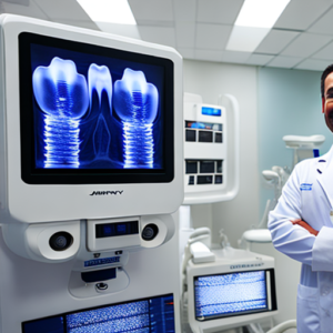
- Cone Beam Computed Tomography (CBCT): Provides three-dimensional imaging, offering unparalleled detail and allowing for precise assessment of bone structure and spatial relationships. CBCT inherently generates higher resolution images than 2D x-rays.
2. Technique – Mastering the Process
Proper technique is arguably more important than equipment alone. Incorrect positioning, patient movement, and improper exposure settings can drastically degrade image quality. Consistent technique standardization is crucial for reliable results.
- Patient Positioning: Precise head position relative to the x-ray beam is paramount. Variations in angulation introduce artifacts and reduce diagnostic accuracy.
- Collimation: Restricting the x-ray field to the area of interest minimizes radiation exposure and improves image sharpness by reducing scatter radiation. Overlapping the collimated field can create severe artifacting.
- Exposure Settings (kVp & mA): Correct kVp (kilovoltage peak) determines the energy of the x-ray beam, influencing contrast resolution. mA (milliampere) controls the quantity of x-rays produced. Optimizing these settings is critical for balancing image quality and patient dose.
- Distance to Film/Sensor: Maintaining consistent distance ensures uniform exposure and reduces magnification artifacts.
3. Patient Factors – A Consideration
Patient factors, although often overlooked, can influence image quality. Patients with motion or poor positioning are more likely to produce blurry images. Small patients, particularly children, often require special techniques due to their limited ability to maintain still positions.
Image Enhancement Techniques for Dental X-Rays
1. Software-Based Enhancements
Many digital radiography systems include built-in software tools designed to enhance image quality post-acquisition. These tools can help reduce noise, improve contrast, and correct minor artifacts.
- Histogram Equalization: Redistributes pixel intensities across the entire range, enhancing contrast where it’s lacking.
- Noise Reduction Filters: Algorithms designed to smooth out random variations in pixel intensity, reducing visual noise. Overuse can lead to loss of detail.
- Contrast Stretching: Expands the dynamic range of pixel intensities, making subtle differences more visible.
2. Post-Processing – Refining the Image
Advanced image processing techniques can further refine dental x-ray images. These often involve specialized software and may require training to utilize effectively.
- Digital Subtraction: Subtracts a previous image (e.g., from a previous day) to highlight changes, aiding in the detection of caries progression or bone loss.
- Bone Density Mapping: Automatically measures bone density based on grayscale values, assisting in the assessment of periodontal disease and other skeletal abnormalities. This is particularly useful for long-term monitoring.
Real-World Examples – The Impact of Image Quality
Case Study 1: Missed Caries Detection
A general dentist routinely used a standard bitewing x-ray technique without adequate collimation. During an annual review, he missed a small carious lesion on the distal surface of tooth number 23. The lesion was only visible after applying a digital enhancement filter that revealed subtle changes in grayscale. The delayed diagnosis resulted in extensive restorative treatment and significant patient anxiety.
Case Study 2: Accurate Periodontal Assessment with CBCT
A periodontist utilized cone beam computed tomography (CBCT) to assess the bone support surrounding teeth in a patient with advanced periodontal disease. The three-dimensional images revealed deep pockets and significant alveolar crestal recession that were not readily apparent on conventional 2D radiographs. This detailed information guided surgical planning and significantly improved treatment outcomes.
Case Study 3: Quantifying Bone Loss
A dentist used bone density mapping software to monitor the progression of osteoporosis in a patient over a five-year period. The software consistently demonstrated an increase in bone loss, prompting earlier intervention with bisphosphonate therapy and ultimately preventing further deterioration.
Conclusion: Investing in Optimal Image Quality
Achieving high-quality dental x-ray images is not simply a matter of convenience; it’s an investment in accurate diagnosis, effective treatment planning, and ultimately, improved patient outcomes. By understanding the factors that influence image quality – from equipment selection to technique execution – dentists can significantly enhance their diagnostic capabilities. Continuous learning and adherence to best practices are paramount in this field.
The evolution of dental imaging technology, particularly CBCT, offers unprecedented detail and precision. However, regardless of the technology employed, mastering fundamental radiographic principles remains essential for producing reliable images that deliver accurate diagnoses. Investing in training and utilizing appropriate enhancement techniques will undoubtedly elevate your diagnostic confidence and contribute to superior patient care.
Key Takeaways
- Equipment Matters: Select the right imaging system for your practice’s needs.
- Technique is Crucial: Consistent, precise technique is paramount to image quality.
- Enhancement Tools are Valuable: Utilize software-based enhancements strategically.
- CBCT Offers Superior Detail: Consider CBCT for complex cases requiring 3D imaging.
- Prioritize Patient Dose: Always aim for the lowest possible radiation dose while maintaining diagnostic quality.
Frequently Asked Questions (FAQs)
- Q: How does digital radiography compare to film radiography? A: Digital radiography offers several advantages, including immediate image viewing, reduced radiation exposure, and the ability to digitally manipulate images.
- Q: What is collimation and why is it important? A: Collimation restricts the x-ray beam to the area of interest, minimizing scatter radiation and improving image sharpness.
- Q: Can I improve image quality by simply taking more pictures? A: While multiple views are necessary for comprehensive diagnosis, taking unnecessary images can increase patient dose and doesn’t necessarily improve image quality if technique is poor.
- Q: What is cone beam computed tomography (CBCT) and when should it be used? A: CBCT provides three-dimensional imaging of the jaws and teeth, ideal for complex cases such as implant planning, surgical guides, and detailed periodontal assessment.