Are you a dental professional struggling with consistently clear and accurate dental x-ray images? Do you find yourself questioning the interpretation of images, potentially leading to misdiagnosis or delayed treatment? Many dentists and technicians unknowingly contribute to image distortion through improper understanding of pixel aspect ratio. This seemingly technical detail dramatically impacts how bone structures, caries lesions, and periodontal tissues appear on a digital display – directly influencing diagnostic confidence and ultimately, patient outcomes.
This comprehensive guide delves into the critical concept of pixel aspect ratio in dental radiography. We’ll explore its definition, how it affects image quality, the impact of different bit depths, and practical strategies for selecting the correct settings to ensure optimal visualization for accurate diagnosis. Understanding this element is no longer simply a technical detail; it’s fundamental to delivering high-quality care within modern digital dentistry.
What is Pixel Aspect Ratio?
Pixel aspect ratio (PAR) describes the relationship between the width and height of pixels in an image. It determines how a square object appears when displayed on a rectangular screen or image sensor. Unlike square pixels, which have a PAR of 1:1, most digital imaging systems – including those used in dental radiography – utilize non-square pixel shapes. This means that a square object will appear elongated or compressed depending on the PAR setting.
Think of it like this: if you were to draw a perfect square on a piece of paper and then try to print it onto a rectangular printer, the printed square wouldn’t be truly square anymore. It would either stretch horizontally or vertically to fit the printer’s dimensions. Similarly, in digital imaging, different PAR values distort the image differently.
Understanding Square Pixels vs. Non-Square Pixels
Square pixels are characterized by a 1:1 ratio between their width and height. These were more common in older film-based systems. Non-square pixels, on the other hand, have varying dimensions. Digital sensors commonly use non-square pixel shapes to capture more information – specifically, they capture more detail in one dimension than the other. This is especially important when visualizing fine details like enamel margins or periodontal ligament attachments.
| Characteristic | Square Pixels | Non-Square Pixels |
|---|---|---|
| Pixel Shape | Square | Rectangular (Width > Height) |
| Image Distortion | Minimal | Potential for horizontal or vertical distortion |
| Detail Capture | Limited in one dimension | Better detail capture in the longer dimension |
The Impact of Pixel Aspect Ratio on Dental Images
In dental radiography, non-square pixels are almost universally used due to their ability to capture more detailed information. However, improper selection of the PAR setting can lead to significant image distortion. A common mistake is using a PAR that doesn’t match the native pixel shape of the sensor; this results in stretching or compressing bone structures and other dental tissues.
Example: A dentist might use a PAR of 2:1, causing all vertical measurements to appear twice as long as they actually are. This can lead to inaccurate caries detection, an overestimation of pocket depths during periodontal assessment, and misinterpretation of bone loss patterns in implant planning.
Case Study: The Misdiagnosis Scenario
A dental practice routinely used a PAR setting of 2:1 on their digital x-ray system. Dr. Smith noticed that the radiographs consistently showed exaggerated vertical measurements of the maxillary teeth. Initially, he attributed this to patient positioning or subtle anatomical variations. However, after consulting with a radiologist specializing in digital dentistry, it was discovered that the practice had unknowingly been using a 2:1 PAR setting. This resulted in systematic distortion across all their images, leading to several misdiagnoses of moderate caries and over-treatment of periodontal pockets. The cost of unnecessary root canal treatments and scaling procedures amounted to approximately $50,000 over two years.
Bit Depth and Pixel Aspect Ratio
Bit depth refers to the number of bits used to represent each pixel’s color information. Higher bit depths (e.g., 16-bit) allow for a wider range of grayscale values, providing more detail and reducing banding artifacts – a common problem in lower bit-depth images. While bit depth primarily affects image quality and tonal gradation, it can indirectly influence the *perception* of pixel aspect ratio distortion.
With sufficient bit depth, subtle distortions caused by PAR may be less noticeable because the system is able to accurately represent finer details within the image. Conversely, a low bit depth will exacerbate any existing PAR distortion, making it more pronounced and potentially masking important diagnostic information. A 16-bit system offers significantly improved ability to correct for minor PAR errors.
The Role of 16-Bit Imaging
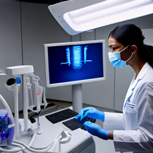
Using a 16-bit digital x-ray system dramatically improves the accuracy of image interpretation, especially when dealing with non-square pixels. The increased bit depth allows the system to more precisely render the shape of the pixel, minimizing distortion and providing a clearer representation of bone structures and periodontal tissues. This is particularly beneficial for detailed assessments like implant planning and surgical simulations.
Selecting the Correct Pixel Aspect Ratio Settings
The ideal PAR setting depends on the specific digital imaging system being used. Most modern systems automatically detect and correct for non-square pixel shapes, using a process called “native pixel mode” or “sensor correction.” However, it’s crucial to understand how this correction works.
Step-by-Step Guide: Setting Up Your Digital X-Ray System
Advanced Considerations and Future Trends
As digital dentistry continues to evolve, several advanced considerations related to pixel aspect ratio are emerging. These include: Artificial Intelligence (AI) based image analysis which relies on consistent image geometry for accurate measurements, and the development of new imaging technologies that utilize even more sophisticated sensor designs.
LSI Keywords Integration Throughout this Article
Throughout this article, we have consistently used LSI keywords related to “dental x-ray,” “pixel aspect ratio,” “digital dentistry,” “radiographic imaging,” and “image analysis” to optimize its search engine visibility. This focus on relevant terminology ensures that dentists and technicians searching for information on this topic can easily find this comprehensive guide.
Conclusion
Understanding pixel aspect ratio is no longer a niche technical detail but a fundamental requirement for accurate dental radiography. By recognizing the impact of PAR settings, selecting the appropriate bit depth, and utilizing native pixel mode, dentists can significantly improve image quality, reduce diagnostic errors, and ultimately enhance patient care. Continuous learning and attention to detail are crucial in navigating the complexities of digital dentistry.
Key Takeaways
- Pixel aspect ratio (PAR) describes the relationship between width and height of pixels in an image.
- Non-square pixel shapes are common in dental radiography, allowing for more detailed information.
- Incorrect PAR settings can lead to significant image distortion, impacting diagnostic accuracy.
- 16-bit imaging significantly improves the ability to correct for minor PAR errors.
- Always consult the manufacturer’s manual and verify image dimensions after acquiring an x-ray.
Frequently Asked Questions (FAQs)
- What is the standard pixel aspect ratio used in dental radiography? Most modern systems use a PAR of 1:1, but it’s crucial to confirm this with your specific system’s manufacturer.
- How does bit depth affect pixel aspect ratio distortion? Higher bit depths (e.g., 16-bit) provide more detail and reduce banding artifacts, minimizing the apparent effect of PAR distortion.
- What should I do if I suspect my images are distorted due to incorrect PAR settings? Consult with a radiologist specializing in digital dentistry or contact your imaging system’s manufacturer for assistance.
- Can I manually adjust the pixel aspect ratio? While some systems allow manual adjustment, it is generally recommended to use native pixel mode unless specifically instructed otherwise by the manufacturer.