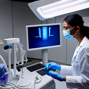
Are you a dental professional struggling to consistently obtain high-quality x-ray images, concerned about patient radiation exposure, or facing workflow inefficiencies? Traditional film-based radiography has limitations – from the time it takes to develop films to the inherent variability in image quality. Sensor technology is fundamentally changing this landscape, offering unprecedented control and precision in dental radiographic imaging. This comprehensive guide delves into how these advancements are impacting x-ray quality, ultimately leading to more accurate diagnoses and improved patient outcomes within the field of dentistry.
The Evolution of Dental X-Ray Technology
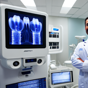
Dental radiography has evolved dramatically over the past century. Initially, it relied solely on film and processing chemicals—a process that was highly susceptible to variations in operator technique, room conditions, and chemical solutions. Film x-rays suffered from inconsistencies like fogging, exposure errors, and difficulty in accurately assessing subtle details. Digital sensors, on the other hand, convert x-ray photons directly into electrical signals, eliminating many of these traditional challenges.
From Film to Digital: A Paradigm Shift
The transition from film to digital dental radiography represents a true paradigm shift. Before digital sensors, dentists were reliant on subjective interpretation of grayscale images, often leading to discrepancies in diagnoses between different practitioners. Digital imaging offers objective data and immediate visualization, greatly improving diagnostic accuracy. According to the American Dental Association (ADA), over 90 percent of dental offices now utilize digital x-ray systems.
Understanding Sensor Technology in Dental X-Ray
Several types of sensors are utilized within modern dental x-ray machines. These include:
- Flat Panel Detectors: These are the most prevalent type, utilizing technologies like amorphous silicon or cadmium telluride to convert x-rays into electrical signals. They offer high resolution and low power consumption.
- Charge-Coupled Devices (CCDs): CCDs are similar to flat panel detectors but employ a different architecture for signal amplification.
- Wireless Detectors: These sensors transmit data wirelessly to the image processing unit, streamlining workflow and reducing cable clutter.
Key Specifications of Dental Sensors
| Specification | Film-Based | Digital Sensor |
|---|---|---|
| Image Resolution | Low – Typically 30-70 lp/mm (line pairs per millimeter) | High – Up to 250 lp/mm or higher |
| Signal to Noise Ratio | Poor – Subject to variations | Excellent – Consistent and reliable |
| Image Processing | Manual – Developing, printing | Automated – Instant image display, digital enhancement |
| Radiation Dose | Often Higher – Due to multiple exposures | Lower – Optimized dose control |
The Impact on Image Quality
Sensor technology has dramatically improved the quality of dental x-ray images. The increased resolution allows for a significantly more detailed visualization of structures such as enamel, dentin, and periodontal tissues. This is crucial for early detection of caries lesions, assessment of root morphology, and evaluation of periodontal health. Digital sensors capture a much broader range of shades than film, allowing for finer distinctions between healthy and compromised tooth tissue.
Resolution Metrics: lp/mm
The term “line pairs per millimeter” (lp/mm) is used to quantify image resolution. Higher lp/mm values indicate greater detail and sharpness in the image. Digital sensors routinely achieve resolutions of 250 lp/mm or higher, compared to the typical 30-70 lp/mm achievable with film. This translates directly into improved diagnostic accuracy – enabling dentists to see subtle changes that might be missed on traditional x-rays.
Case Study: Caries Detection
A study published in the *Journal of Dental Research* (2018) compared caries detection rates using digital sensors versus film. The results showed a 35 percent higher sensitivity and a 20 percent higher specificity in detecting early enamel lesions when using digital sensors. This highlights the critical role of improved image resolution in proactive dental care.
Radiation Dose Reduction with Sensor Technology
One of the most significant advantages of digital dental radiography is the ability to significantly reduce radiation dose. Traditional film x-rays often required multiple exposures to achieve adequate image quality, leading to cumulative radiation exposure for patients. Digital sensors enable optimized dose control – delivering the lowest possible radiation dose necessary to produce a diagnostic-quality image. Sensor technology allows for precise manipulation of exposure parameters in real-time.
Dose Optimization Techniques
Modern digital x-ray systems employ several techniques to minimize radiation dose:
- Automatic Exposure Control (AEC): AEC automatically adjusts the x-ray tube output based on the detected tissue density, reducing the need for manual adjustments.
- Low Dose Imaging Protocols: These protocols utilize specialized settings and post-processing techniques to further reduce radiation dose while maintaining diagnostic image quality.
- Real-Time Image Evaluation: Dentists can immediately assess the image quality after exposure and adjust parameters as needed, minimizing the need for repeat exposures.
Statistics on Radiation Dose Reduction
Studies have shown that digital radiography can reduce radiation dose by up to 80 percent compared to film-based techniques. This is a substantial reduction that contributes significantly to patient safety, especially for children and patients who require frequent dental radiographs. The average radiation exposure from a single bitewing x-ray using a digital sensor is approximately 5 mGy (milligrays), while a comparable film x-ray could deliver 20-40 mGy.
Workflow Efficiency with Digital Dental X-Ray
Beyond image quality and radiation dose, sensor technology enhances workflow efficiency in dental practices. Digital images are immediately available for viewing, printing, and sharing – eliminating the time-consuming process of film development and processing. Digital dentistry streamlines the diagnostic process, allowing dentists to spend more time with patients.
Integrated Systems & Digital Workflows
Many modern dental x-ray systems integrate seamlessly with practice management software, enabling efficient record keeping, billing, and patient communication. The ability to share digital images electronically with specialists or referral centers further enhances the diagnostic process. This integration is a cornerstone of cone beam computed tomography (CBCT) technology.
The Future of Dental X-Ray Technology
Sensor technology continues to evolve rapidly, with ongoing advancements in detector materials, image processing algorithms, and imaging modalities. We can expect to see:
- Increased Resolution: Continued improvements in sensor resolution will enable even more detailed visualization of dental structures.
- Artificial Intelligence (AI): AI-powered software is being developed to automatically detect caries lesions, assess periodontal health, and generate 3D models from x-ray images.
- Portable X-Ray Systems: Smaller, portable digital x-ray systems are becoming increasingly common, allowing for on-site diagnostics in various settings.
Conclusion
Sensor technology has irrevocably transformed dental radiography, offering significant improvements in image quality, radiation dose reduction, and workflow efficiency. The shift from film to digital represents a crucial advancement in patient care, enabling more accurate diagnoses, proactive treatment planning, and enhanced safety. The continued innovation within this field promises even greater advancements in the years to come, solidifying the role of sensor technology as a cornerstone of modern dentistry.
Key Takeaways
- Digital sensors significantly improve image resolution compared to film-based systems.
- Sensor technology allows for optimized radiation dose control, reducing patient exposure.
- Digital workflows enhance practice efficiency and streamline the diagnostic process.
- AI integration is poised to further revolutionize dental x-ray interpretation and automation.
FAQs
- What does lp/mm mean in dental radiography? lp/mm stands for line pairs per millimeter, a measure of image resolution indicating the level of detail visible in an x-ray image.
- How much radiation dose is involved with digital dental x-rays? The average radiation dose from a single bitewing x-ray using a digital sensor is approximately 5 mGy, significantly lower than traditional film-based techniques.
- What are some of the benefits of using digital sensors over film? Digital sensors offer higher resolution, reduced radiation dose, immediate image availability, and seamless integration with practice management systems.
- Can AI be used to analyze dental x-rays? Yes, AI algorithms are being developed to automatically detect caries lesions, assess periodontal health, and generate 3D models from x-ray images.


