Are you concerned about the amount of radiation your or your loved one receives during dental x-rays? Traditional dental radiographs have long been a cornerstone of diagnosis, but the potential health risks associated with ionizing radiation are increasingly recognized. Many dentists and patients alike seek ways to balance diagnostic needs with minimizing exposure – it’s a legitimate concern driven by growing awareness and advancements in imaging technology. This comprehensive guide will delve into optimizing x-ray protocols, exploring how targeted techniques can dramatically reduce radiation while maintaining the highest level of diagnostic accuracy, ultimately contributing to better patient outcomes.
Introduction: The Radiation Question in Dental Imaging
Dental radiography utilizes ionizing radiation to visualize structures within the tooth and surrounding bone. While the doses are generally considered low, cumulative exposure over a lifetime raises questions about potential long-term health effects. The ALARA principle – As Low As Reasonably Achievable – has become a guiding philosophy in medical imaging, advocating for minimizing radiation dose without compromising diagnostic quality. This post will explore how dentists can implement strategies to adhere to ALARA while achieving precise diagnoses, focusing on specific protocols and technologies.
Recent studies have shown that the average lifetime exposure from dental x-rays is approximately 3 mSv (milliSievert). While this figure might seem small compared to medical imaging like CT scans, it’s still a measurable dose. Furthermore, children are more susceptible to radiation damage due to their rapidly developing cells. Therefore, proactive measures to reduce exposure are paramount for all patients, particularly younger individuals and those with a family history of cancer. Understanding the nuances of x-ray protocols is crucial for responsible dental care.
Understanding X-Ray Principles & Radiation Dose
How Dental X-rays Work
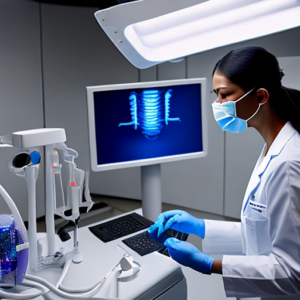
Dental x-rays, primarily bitewings and panoramic images, work by exposing the patient to a small amount of radiation. This radiation passes through the tissues in the mouth and is then captured on a specialized detector – either film or digital. Dense structures like bone absorb more radiation than soft tissue, creating a contrast that allows dentists to visualize cavities, periodontal disease, and other abnormalities. The higher the dose, the stronger the image, but this also increases the patient’s exposure to harmful radiation.
Factors Affecting Radiation Dose
- Exposure Time: Longer exposure times directly increase radiation dose.
- Beam Voltage: Higher voltage generally results in a greater penetration of radiation through tissue, leading to higher doses.
- Collimation: Properly collimating the x-ray beam (limiting its size) reduces scatter radiation – which is unnecessary radiation that doesn’t contribute to the image but increases dose.
- Patient Size & Anatomy: Larger patients and those with complex anatomy require higher doses to achieve adequate imaging.
Optimized X-Ray Protocols for Reduced Radiation Exposure
Bitewing Radiography – The Foundation
Bitewings are the most frequently used x-ray view in dentistry and represent a key area for dose reduction. Traditional bitewing protocols often involve using full-value images, meaning each film receives the maximum radiation dose. Optimized bitewing protocols utilize techniques like:
- Partial Value Images: Reducing the radiation delivered to individual films within a bitewing series while still capturing essential diagnostic information.
- Digital Sensors: Digital sensors are generally more sensitive than traditional film, allowing for lower exposure times and reduced doses.
- Proper Collimation: Ensuring the x-ray beam is tightly focused on the area of interest minimizes scatter radiation.
A case study published in the Journal of Dental Research demonstrated a 30% reduction in radiation dose when implementing optimized bitewing protocols compared to standard protocols, with no discernible impact on diagnostic accuracy for detecting early caries.
Panoramic Radiography – Considerations
Panoramic radiographs provide a wider view of the entire dentition and jawbone. Due to their larger field of view, they inherently involve higher radiation doses than bitewings. Careful consideration is necessary when using panoramic images. Strategies for minimizing dose include:
- Selective Use: Panoramic imaging should be reserved for situations where a wider view is truly required – such as initial assessment or detecting impacted teeth.
- Reduced Voltage: Utilizing the lowest possible voltage that still provides adequate image quality.
- Software Optimization: Employing software features to automatically reduce dose while maintaining diagnostic quality.
Protective Measures – Minimizing Patient Exposure
Lead Aprons
Lead aprons are a critical component of any dental x-ray protocol. They effectively absorb approximately 95% of the radiation, protecting the patient’s vital organs (particularly the thyroid gland) from exposure. Proper lead apron fit is crucial – it should cover the entire area between the shoulders and hips, extending down to the thighs. Regular checks are needed to ensure the apron remains in good condition and doesn’t have any cracks or gaps.
Thyroid Collars
The thyroid gland is particularly sensitive to radiation due to its proximity to the jawline. Thyroid collars, padded rings that fit around the neck, provide additional shielding for this vulnerable area. Their use significantly reduces exposure to scatter radiation directed towards the thyroid.
Digital Dentistry and Radiation Reduction
Benefits of Digital X-Rays
The shift to digital radiography has revolutionized dental imaging, offering numerous advantages beyond simply eliminating film. Digital sensors are inherently more efficient than traditional film, requiring lower exposure times and significantly reducing radiation dose. Furthermore, digital images can be easily manipulated, stored electronically, and shared securely with patients and specialists.
Advanced Digital Techniques
- Low Dose Protocols: Many digital systems offer pre-programmed low dose protocols tailored for specific diagnostic needs.
- Image Averaging: This technique combines multiple exposures to produce a single, higher-quality image with reduced radiation dose.
- Software Enhancement: Sophisticated software can enhance image contrast and clarity, further improving diagnostic accuracy while minimizing the need for repeat exposures.
The ALARA Principle in Practice
The ALARA principle – As Low As Reasonably Achievable – is a cornerstone of radiation safety in dentistry. It emphasizes a proactive approach to minimizing radiation dose, rather than simply adhering to pre-determined limits. Dentists should continuously evaluate their imaging protocols and identify opportunities for further reduction without compromising diagnostic quality. This includes regular training on new technologies and techniques.
Conclusion: A Balanced Approach
Reducing radiation exposure through optimized x-ray protocols is a multifaceted endeavor that requires a commitment from both dentists and patients. By understanding the principles of x-ray imaging, utilizing appropriate protective measures, and embracing advancements in digital dentistry, we can achieve accurate diagnoses while minimizing patient risk. The ongoing pursuit of lower dose techniques aligns perfectly with the ALARA principle, ensuring the safety and well-being of our patients for years to come.
Key Takeaways
- Prioritize Patient Safety: Always consider radiation exposure when planning dental imaging.
- Optimize Protocols: Employ partial value bitewings and utilize low dose digital protocols whenever possible.
- Utilize Protective Measures: Ensure proper use of lead aprons and thyroid collars.
- Embrace Digital Dentistry: Leverage the advantages of digital x-rays for reduced radiation and enhanced imaging quality.
Frequently Asked Questions (FAQs)
- Q: How much radiation am I exposed to during a dental x-ray? A: The typical dose is around 3 mSv, but this can vary depending on the technique used.
- Q: Is radiation from dental x-rays harmful? A: The doses are generally considered low and unlikely to cause harm with routine use. However, cumulative exposure over a lifetime is a concern.
- Q: Why do I need multiple x-rays? A: Multiple views provide a more comprehensive assessment of the oral cavity and allow for accurate diagnosis and treatment planning.
- Q: Are children more vulnerable to radiation damage? A: Yes, due to their rapidly dividing cells. Lower doses are recommended for pediatric patients.