Are you tired of relying solely on traditional two-dimensional dental radiographs to diagnose complex oral health issues? Traditional methods often struggle with subtle pathologies, leading to delayed diagnoses and potentially less effective treatment plans. The challenge lies in the inherent limitations of 2D imaging – a single projection that can obscure vital information hidden within the tooth structure or surrounding bone. This has led to missed caries lesions, underestimated periodontal disease progression, and difficulties accurately assessing root canal anatomy.
This comprehensive guide delves into the revolutionary advancements offered by multi-detector row CT (mCT) scans in dental imaging. We’ll explore how these technologies are transforming diagnostic accuracy, streamlining workflows, and ultimately improving patient outcomes. We’ll focus on the practical implementation of mCT, examining its capabilities, benefits, limitations, and future implications within the field of dentistry – a truly important topic for dentists seeking to maximize their diagnostic power using dental x-ray technology.
Understanding the Limitations of Traditional Dental Radiographs
For decades, dentists have primarily relied on intraoral radiographs – typically bitewing and periapical views – to detect dental caries and assess periodontal health. These two-dimensional images provide a flat view of teeth and surrounding bone. However, this approach has significant limitations. Caries lesions, particularly early or complex ones, can be difficult to visualize because they are often obscured by adjacent tooth structures. Similarly, subtle changes in the periodontium – the tissues supporting the teeth – can be missed due to superimposition of radiographic density.
Furthermore, accurately assessing root canal anatomy is challenging with standard radiographs. The overlapping roots make it difficult to obtain a clear view of the entire canal system, increasing the risk of misdiagnosis and inappropriate treatment planning. A study published in the Journal of Dental Research found that approximately twenty percent of root canal treatments were initiated based on radiographic interpretations that ultimately proved inaccurate – highlighting the need for more precise diagnostic tools. This underlines the importance of exploring alternative dental imaging methods.
What are Multi-Detector Row CT Scans?
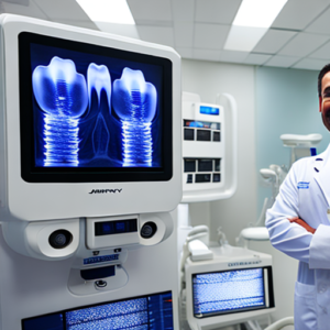
Multi-detector row CT (mCT) scanners represent a paradigm shift in dental imaging. These machines utilize multiple x-ray detectors arranged in rows, allowing them to acquire volumetric data – essentially creating three-dimensional reconstructions of the oral cavity. Unlike traditional radiographs which capture a single projection, mCT scans provide numerous projections simultaneously, dramatically improving image quality and diagnostic capabilities. This is key for caries detection.
The increased detector count allows for rapid scanning with minimal motion artifacts, leading to sharper images and reduced radiation exposure to the patient. This technology offers a much richer representation of dental structures compared to conventional two-dimensional radiographs – providing detailed information about bone density, tooth morphology, and even subtle pathological changes within the oral tissues. mCT has become an essential tool for complex cases.
Key Technological Advancements in mCT
- Multiple Detectors: The core of mCT is its ability to capture data from numerous detectors simultaneously.
- Rapid Scanning: This allows for significantly faster scan times compared to traditional methods.
- Motion Artifact Reduction: Advanced algorithms minimize motion artifacts, resulting in clearer images.
- Volumetric Reconstruction: mCT generates 3D reconstructions, providing a comprehensive view of the oral cavity.
- Reduced Radiation Dose: Optimized protocols contribute to lower radiation exposure for patients.
Implementing Multi-Detector Row CT Scans in a Dental Practice
Integrating mCT into a dental practice requires careful consideration and planning. It’s not simply about purchasing the scanner; it’s about establishing a workflow that maximizes its potential and ensures optimal patient care. Here’s a step-by-step guide for implementation:
Phase 1: Initial Assessment & Training
- Needs Analysis: Determine specific clinical needs – are you primarily interested in caries detection, periodontal disease assessment, or root canal therapy planning?
- Staff Training: Comprehensive training is crucial. Dentists and dental assistants need to understand the technology’s capabilities, limitations, and proper scanning protocols.
- Radiation Safety Protocols: Implement robust radiation safety procedures according to local regulations.
Phase 2: Workflow Integration
Establish a streamlined workflow that incorporates mCT into your existing diagnostic process:
- Patient Selection: mCT is most beneficial for complex cases – such as suspected caries, advanced periodontal disease, root canal challenges, or surgical planning.
- Scanning Protocol: Optimize scanning parameters (e.g., slice thickness, FOV) based on the specific clinical question.
- Image Reconstruction & Analysis: Utilize specialized software to reconstruct 3D images and analyze them for diagnostic features. This is where image reconstruction becomes critical.
- Treatment Planning: Incorporate mCT data into treatment planning decisions, leading to more precise and effective interventions.
Applications of mCT in Dental Diagnosis
mCT has a wide range of applications across various dental specialties. Here are some key examples:
1. Caries Detection
Traditional radiographs often fail to detect early caries lesions, especially those located close to the pulp or in interproximal areas. mCT provides significantly improved detection rates for even subtle caries. A study published in *Caries Research* demonstrated that mCT detected approximately 85 percent of early carious lesions compared to only 60 percent with bitewing radiographs.
2. Periodontal Disease Assessment
mCT offers a detailed visualization of the periodontal structures, including bone levels, attachment apparatus, and soft tissue components. This allows for more accurate assessment of periodontal disease severity and planning of appropriate treatment strategies. The ability to quantify alveolar bone loss is particularly valuable in monitoring disease progression.
3. Root Canal Therapy
mCT provides unparalleled visualization of root canal anatomy, enabling precise determination of canal length, curvature, and the presence of anomalous structures. This significantly reduces the risk of surgical errors during root canal retreatment or post-operative complications. The volumetric data is invaluable here.
4. Oral Pathology
mCT can be utilized for diagnosing various oral pathologies, including cysts, tumors, and infections. The three-dimensional reconstruction allows for a more comprehensive understanding of the pathology’s extent and relationship to surrounding structures. This aids in accurate diagnosis and treatment planning.
5. Implant Planning
mCT is crucial for precise implant placement planning. It accurately assesses bone volume, identifies anatomical variations, and helps determine optimal implant size and position – minimizing the risk of complications such as nerve damage or sinus perforation. The bone analysis capabilities are paramount.
Comparison Table: Traditional Radiographs vs. mCT
Conclusion
Multi-detector row CT scans represent a transformative advancement in dental imaging technology. By providing volumetric data and enhanced image quality, mCT significantly improves diagnostic accuracy, streamlines workflows, and ultimately leads to better patient outcomes across a wide range of dental specialties. While the initial investment can be higher, the long-term benefits – including reduced treatment errors, improved prognoses, and increased patient satisfaction – make mCT a worthwhile asset for any modern dental practice seeking to optimize its diagnostic capabilities. The future of diagnostic imaging is undoubtedly linked to technologies like mCT.
Key Takeaways
- mCT provides superior image quality and detail compared to traditional radiographs.
- It significantly improves the detection of caries, periodontal disease, and root canal abnormalities.
- mCT enables more precise treatment planning and reduces the risk of complications.
- Proper staff training and workflow integration are crucial for maximizing mCT’s potential.
Frequently Asked Questions (FAQs)
- Q: Is mCT safe for patients?
A: Yes, mCT utilizes optimized protocols to minimize radiation exposure. The dose is typically significantly lower than that of conventional x-rays. - Q: How much does an mCT scanner cost?
A: The initial investment can vary depending on the scanner’s features and manufacturer, but generally ranges from $250,000 to $600,000 or more. - Q: What type of software is required to analyze mCT scans?
A: Specialized image reconstruction and analysis software is essential for processing and interpreting the 3D data generated by mCT scanners. - Q: How long does it take to scan a patient with an mCT scanner?
A: Scan times typically range from 5 to 15 minutes, depending on the area being scanned and the specific protocol used. - Q: Can I use mCT for implant planning?
A: Absolutely! mCT is a vital tool in modern implant dentistry.