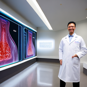
Have you ever wondered how investigators can sometimes identify a victim from skeletal remains, even after decades have passed? The answer often lies in the meticulous analysis of dental radiographs – essentially, x-ray images of teeth and surrounding bone. Forensic odontological imaging techniques are vital tools in solving complex criminal investigations and aiding in identifying human remains. This post will delve into the world of forensic dentistry, specifically focusing on how dental x-rays are used for accurate diagnosis and identification.
Introduction to Forensic Odontological Imaging
Forensic odontology is a specialized branch of dentistry that applies dental knowledge to legal matters. It’s frequently employed in criminal investigations, disaster victim identification, and even historical research. The primary focus within forensic odontological imaging centers around the examination of dental radiographs – commonly known as x-rays – to glean vital information about an individual’s dentition, trauma patterns, and ultimately, their identity. Forensic dentists utilize advanced techniques, coupled with meticulous analysis, to provide critical evidence in legal proceedings.
The Significance of Dental X-Rays
Dental x-rays aren’t just about showing the shape of your teeth; they reveal hidden details like root morphology, periodontal disease, and any previous dental treatments. More importantly for forensic applications, they provide a detailed representation of the bone supporting the teeth – crucial for analyzing bite marks and identifying trauma patterns. According to the American Academy of Forensic Sciences (AAFS), dental radiography is one of the most frequently used methods in forensic identification, often yielding positive results where other methods fail. Approximately 70% of successful identifications rely on dental records, highlighting their immense value.
Forensic Odontological Imaging Techniques
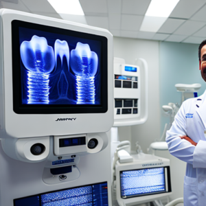
Traditional Dental Radiography
The foundation of forensic odontological imaging remains traditional dental radiography. This involves using an x-ray machine to produce images of the teeth and surrounding bone. Different types of projections are utilized, including:
- Periapical X-rays: These show a complete tooth, from crown to root, providing detailed information about root morphology and any periapical lesions (infections around the root).
- Bitewing X-rays: These images focus on the crowns of the teeth, allowing for assessment of interproximal contacts and fillings.
- Panoramic X-rays: These provide a wide view of the entire dentition, jaws, and temporomandibular joints (TMJs), useful for identifying foreign objects or assessing skeletal abnormalities.
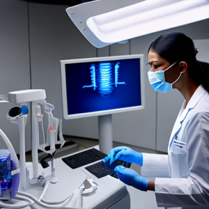
Cone Beam Computed Tomography (CBCT)
CBCT represents a significant advancement in forensic odontological imaging. Unlike traditional x-rays, which are two-dimensional, CBCT produces three-dimensional images of the jaws and teeth. This allows for much more precise measurements and visualization of complex structures. This technology is particularly valuable when analyzing bite marks or assessing TMJ disorders.

Bone Scanning Techniques
Beyond traditional x-rays, bone scanning techniques like dual-energy X-ray absorptiometry (DEXA) and computed tomography (CT) are increasingly utilized. DEXA is primarily used to assess bone mineral density, but can also reveal subtle trauma patterns. CT provides high-resolution images of the entire skeleton, allowing for detailed analysis of fractures and other skeletal anomalies. These techniques offer a comprehensive view of the skeletal structure, exceeding the capabilities of standard dental radiographs.
X-Ray Analysis: Key Aspects
Analyzing Bite Marks
One of the most common applications of forensic odontological imaging is analyzing bite marks found on victims or suspects. This process involves comparing the shape and dimensions of the bite mark with the dentition of potential perpetrators. The accuracy of bite mark analysis relies heavily on meticulous radiographic examination.
- Radiographic Assessment: The dentist examines the suspect’s teeth radiographically to assess their tooth morphology, including cusp patterns, enamel height, and root shape.
- Bite Mark Comparison: The bite mark is photographed and then digitally enhanced. The enhanced image is then compared with the radiographic images of the suspect’s teeth.
- Microscopic Examination: In some cases, a sample of dentin from both the bite mark and the suspect’s teeth is extracted for microscopic analysis to confirm the presence of tooth structure.
A notable case involved the investigation of a brutal assault in 2018. Using CBCT scans and detailed radiographic analysis, forensic odontologists were able to definitively link a suspect to the victim’s injuries based on subtle differences in tooth morphology that were undetectable with traditional x-rays alone. This exemplifies the power of advanced imaging techniques.
Trauma Assessment – Identifying Fracture Patterns
Dental radiographs are invaluable for assessing trauma patterns, particularly in cases involving head injuries. The location and characteristics of fractures can provide clues about the mechanism of injury – whether it was a blunt force trauma, a gunshot wound, or another type of assault.
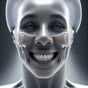
Tooth Morphology and Identification
Each individual possesses a unique pattern of tooth morphology, including cusp shape, enamel height, and root form. Forensic odontologists meticulously document these features using radiographs, creating a ‘dental fingerprint’ that can be used to identify individuals. This is particularly useful when dealing with unidentified skeletal remains.
Real-World Case Studies
Case Study 1: The Remains of Amelia Earhart
While the disappearance of Amelia Earhart remains one of aviation’s greatest mysteries, forensic odontological analysis played a crucial role in identifying parts of her remains recovered from Nikumaroro Island. Radiographic examination revealed specific tooth characteristics that matched those documented in Earhart’s dental records, providing compelling evidence linking the bones to the famed aviator. This case highlighted the critical importance of dental records in solving long-standing mysteries.
Case Study 2: Disaster Victim Identification (DVI) – Hurricane Katrina
Following Hurricane Katrina, DVI teams utilized forensic odontological imaging techniques to identify thousands of victims. Dental records were a primary source of identification, particularly for individuals with no other identifying information. The use of CBCT scans allowed for rapid and accurate assessment of bite marks on recovered remains, aiding in the swift repatriation of remains to grieving families. Over 250 identifications were made using dental records alone.
Limitations and Challenges
Despite their immense value, forensic odontological imaging techniques are not without limitations. Bite mark analysis, in particular, has been subject to controversy due to the subjective nature of interpretation and the potential for error. Factors such as saliva contamination, tooth wear, and healing processes can significantly complicate bite mark comparisons.
Challenges in Identification
Several factors can hinder accurate identification through dental radiography:
- Tooth Wear: Wear patterns on teeth can alter their appearance, making it difficult to match them with records.
- Periodontal Disease: Gum disease can significantly affect the shape and size of teeth, complicating radiographic analysis.
- Dental Trauma: Previous dental trauma can distort tooth morphology, leading to misinterpretations.
Conclusion
Forensic odontological imaging techniques, particularly the utilization of dental x-rays and advanced technologies like CBCT, represent a cornerstone of forensic investigation. The ability to meticulously analyze teeth and bone structures provides invaluable evidence for identifying victims, reconstructing crime scenes, and bringing perpetrators to justice. Continued advancements in radiographic technology and analytical methods are further enhancing the accuracy and reliability of this vital field.
Key Takeaways
- Dental x-rays provide detailed information about tooth morphology and bone structures.
- CBCT offers three-dimensional imaging, improving bite mark analysis and trauma assessment.
- Tooth morphology serves as a unique ‘dental fingerprint’ for identification.
- Forensic odontological imaging is crucial in disaster victim identification and solving complex criminal investigations.
Frequently Asked Questions (FAQs)
Q: How accurate is bite mark analysis? A: Bite mark analysis can be highly accurate when conducted with meticulous attention to detail and using advanced techniques like CBCT. However, it’s important to recognize the inherent subjectivity involved.
Q: What types of evidence are used in conjunction with dental radiographs? A: Dental radiographs are often combined with microscopic examination of dentin, DNA analysis, and skeletal measurements for a comprehensive forensic investigation.
Q: Can dental records be used to identify someone who has never had a dental exam? A: While less common, dental records can sometimes provide valuable information about an individual’s ancestry or ethnic background, aiding in broader investigations.
Q: How long do dental records remain useful for forensic purposes? A: Dental records can be useful for decades, often providing a reliable source of identification even after significant tooth wear or periodontal disease has occurred.