Are you a dentist, dental student, or anyone involved in oral healthcare struggling to interpret the nuances hidden within dental x-ray images? Traditional dental radiographs often struggle to clearly visualize subtle abnormalities, leading to delayed diagnoses and potentially less effective treatment plans. Many conditions like early caries, periodontal disease, and even complex root canal issues can be obscured by bone density, making accurate assessment challenging. This article delves into how contrast agents revolutionize the process of decoding these images, providing unparalleled clarity for improved diagnostic accuracy and ultimately, better patient outcomes. Understanding this technology is fundamental to modern dental practice.
Introduction: The Challenge of Traditional Dental Radiography
Dental radiographs, or x-rays, have been a cornerstone of oral healthcare for over a century. However, the inherent limitations of standard techniques – primarily relying on variations in bone density to identify pathology – frequently result in missed diagnoses. Bone is naturally much denser than soft tissues like tooth enamel and periodontal ligament, creating a significant contrast that can mask early signs of disease. For example, a small, early caries lesion might appear as a faint shadow within the dense bone structure, making it difficult for even experienced dentists to detect without specialized tools or techniques. This often leads to delayed treatment initiation and potentially more complex restorative procedures later on.
What are Contrast Agents in Dental X-Ray?
Contrast agents are substances administered to a patient before radiographic imaging that temporarily alter the density of tissues, making them more visible on x-ray images. Essentially, they enhance contrast between different structures, allowing for clearer visualization of abnormalities. In dentistry, this is particularly crucial because bone often dominates the dental x-ray image, obscuring smaller pathologies within the tooth itself or surrounding soft tissues.
The principle behind their effectiveness lies in their ability to absorb X-rays differently than the surrounding tissue. This difference in absorption creates a brighter area on the film or digital sensor, highlighting the presence of the agent and, consequently, the underlying structure it’s associated with. Without contrast agents, many subtle dental conditions would remain hidden, significantly impacting diagnostic accuracy.
Types of Contrast Agents Used in Dentistry
Several types of contrast agents are utilized in dentistry, each with its own advantages and considerations. These include:
- Ionic Radiopaque Compounds: These compounds, such as barium sulfate, are the most commonly used. They create a dense, opaque area on the x-ray image by absorbing X-rays intensely. Barium is frequently employed for visualizing the periodontal ligament space and detecting caries.
- Amido-Iodinated Contrast Agents: These agents, like iohexol and metrizamide, are non-ionic and have a higher water content than barium sulfate. They offer better radiopacity and are less likely to cause adverse reactions. They are often preferred for complex procedures requiring detailed visualization.
- Gadolinium-Based Contrast Agents: While less frequently used in routine dental x-rays due to concerns about gadolinium retention, they can be employed in specialized applications like assessing bone metabolism or diagnosing certain oral pathologies.
Applications of Contrast Agents in Dental X-Ray Visualization
Contrast agents are utilized across a wide range of dental procedures and diagnostic scenarios. Their ability to dramatically improve image clarity has revolutionized several areas of dentistry. Here are some key examples:
1. Caries Detection
Early caries detection is paramount for successful treatment. Traditional radiographs often fail to reveal the early stages of decay, particularly when located in proximity to bone. Using a contrast agent like barium sulfate can highlight the small cavities forming within enamel or dentin, allowing dentists to intervene before significant damage occurs. Studies have shown that the use of contrast agents significantly improves caries detection rates compared to standard radiography, especially in areas adjacent to bone.
2. Periodontal Disease Assessment
Contrast agents are invaluable for assessing periodontal disease. They effectively visualize the periodontal ligament space – the area separating the tooth root from surrounding bone – which is often difficult to discern on regular radiographs. This allows dentists to accurately assess bone loss, inflammation, and other signs of periodontal breakdown. A 2018 study published in the Journal of Periodontology demonstrated that using iohexol significantly improved the detection of pocket depth and probing attachment level compared to non-contrast imaging.
3. Root Canal Visualization
During root canal treatment, contrast agents can be used to improve visualization of the root canals themselves, particularly when they are narrow or curved. This aids in accurate instrumentation and irrigation, minimizing the risk of complications. In cases where the canal is obscured by bone, a small amount of contrast agent administered intravenously can dramatically enhance visibility.
4. Dental Implant Assessment
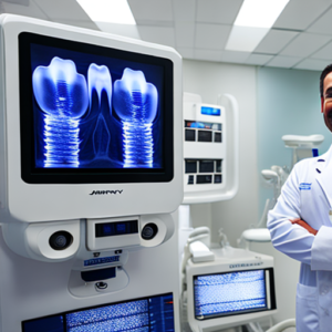
When evaluating dental implants, contrast agents help assess bone integration around the implant’s neck. Detecting marginal bone loss or peri-implantitis early on is crucial for maintaining long-term implant success. Using iohexol in conjunction with cone-beam computed tomography (CBCT) provides a detailed assessment of this critical area.
5. Oral Pathology Diagnosis
Contrast agents can be utilized to detect and analyze oral tumors, cysts, or other lesions that might otherwise be difficult to visualize on standard radiographs. They assist in characterizing the lesion’s borders, size, and relationship to surrounding structures.
Step-by-Step Guide: Using Contrast Agents for Caries Detection
Here’s a simplified guide to using contrast agents like barium sulfate for caries detection:
- Patient Preparation: The patient is given a small, measured dose of the contrast agent (typically via oral administration).
- Radiographic Acquisition: A periapical or bitewing x-ray is taken immediately after administering the contrast agent.
- Image Interpretation: The dentist carefully analyzes the image for any increased radiopacity within the tooth structure, indicating a potential caries lesion.
Comparison Table: Contrast Agents vs. Standard Dental Radiography
Conclusion
Contrast agents have fundamentally transformed the landscape of dental x-ray visualization. Their ability to enhance tissue contrast has dramatically improved diagnostic accuracy, leading to earlier detection and more effective treatment planning for a wide range of oral conditions. Continued advancements in contrast agent technology and imaging techniques will undoubtedly further refine our ability to decode dental x-ray images and ultimately improve patient care. The strategic use of these agents is no longer an optional step but an essential component of modern dental practice.
Key Takeaways
- Contrast agents dramatically improve image clarity in dental radiographs, making it easier to detect subtle abnormalities.
- They are used across various applications, including caries detection, periodontal disease assessment, root canal treatment, and implant evaluation.
- Different types of contrast agents exist, each with specific properties and considerations.
- Proper utilization of contrast agents significantly enhances diagnostic accuracy and contributes to better patient outcomes.
Frequently Asked Questions (FAQs)
Q: Are contrast agents safe for all patients?
A: Generally, yes. However, individuals with allergies or kidney problems should consult their physician before receiving a dental x-ray with a contrast agent.
Q: How long does it take for a contrast agent to leave the body?
A: The elimination time varies depending on the specific agent used, but most are eliminated within 24 to 72 hours.
Q: What happens if a patient has an allergic reaction to a contrast agent?
A: Immediate medical attention is required. The dentist should have protocols in place for managing allergic reactions, including epinephrine administration if necessary.
Q: Can children receive contrast agents?
A: Yes, but only under the guidance of a qualified dentist or physician. The dosage and administration method are carefully adjusted for pediatric patients.
Q: What is the role of CBCT in conjunction with contrast agents?
A: Cone-beam computed tomography (CBCT) provides 3D imaging, which can be greatly enhanced by using contrast agents to visualize fine details within complex structures like roots and implants. This offers a more comprehensive diagnostic view than traditional 2D x-rays.


